Home » Without Label » Diagram Of Hip.and Back.muscles / Female Torso Musculature Labelled Back Muscles Anatomy Anatomy Of Muscles Hip And Lower Back Human Muscle Anatomy Body Anatomy Shoulder Muscle Anatomy / Each of the muscles diagrams illustrates a slightly different set of muscles.
Diagram Of Hip.and Back.muscles / Female Torso Musculature Labelled Back Muscles Anatomy Anatomy Of Muscles Hip And Lower Back Human Muscle Anatomy Body Anatomy Shoulder Muscle Anatomy / Each of the muscles diagrams illustrates a slightly different set of muscles.
Diagram Of Hip.and Back.muscles / Female Torso Musculature Labelled Back Muscles Anatomy Anatomy Of Muscles Hip And Lower Back Human Muscle Anatomy Body Anatomy Shoulder Muscle Anatomy / Each of the muscles diagrams illustrates a slightly different set of muscles.. Want to learn more about it? They are the biceps femoris (long head and short head), semimembranosus, and semitendinosus. They begin under the gluteus maximus behind the hip bone and attach to the tibia at the knee. Most modern anatomists define 17 of these muscles, although some additional muscles may sometimes be considered. Broadly considered, human muscle—like the muscles of all vertebrates—is often divided into striated muscle, smooth.
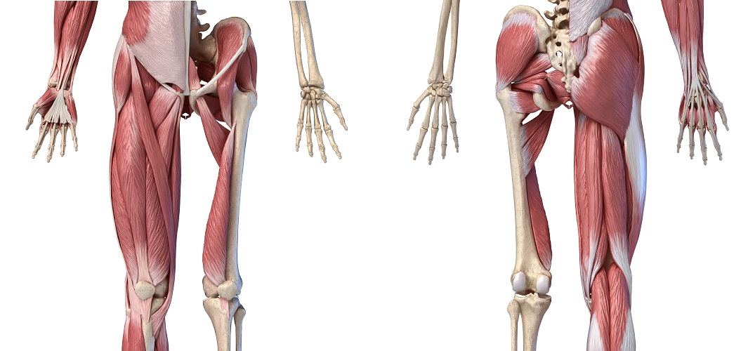
In the back of the thigh, the hamstring muscles affect hip and knee movement. The core muscles are those in the abdomen, back, and pelvis, and they also stabilize the body and assist in tasks, such as lifting weights. Common hip and back pain causes include injury to muscles from overuse disc injurydegeneration or spinal stenosis. The hip muscle diagram below shows a number of the muscles we will be discussing in the next sections. Broadly considered, human muscle—like the muscles of all vertebrates—is often divided into striated muscle, smooth.
Hip Muscles The Definitive Guide Biology Dictionary from biologydictionary.net Muscles of hip bone and spine. They are the biceps femoris (long head and short head), semimembranosus, and semitendinosus. Francesca salvador msc last + show all. All of these things can lead to long term back pain (and chronic complaining!). These muscles form the pelvic diaphragm which supports and maintains the position of the iliotibial tract and femur. It joins the lower limb to the pelvic girdle. The hip muscle diagram below shows a number of the muscles we will be discussing in the next sections. Muscles of the posterior … category:
In the back of the thigh, the hamstring muscles affect hip and knee movement.
Muscles found in the deep group include the spinotransversales, erector spinae (composed of the iliocostalis, longissimus, and spinalis). Francesca salvador msc last + show all. The human back extends from the buttocks to the posterior portion of the neck and shoulders. Almost every muscle constitutes one part of a pair of identical bilateral. The main muscles of the hip and pelvis consistsof the iliopsoas, pectinues, rectus femoris and sartorius at the front. Other muscles are small and cover much less space. They begin under the gluteus maximus behind the hip bone and attach to the tibia at the knee. Want to learn more about it? The muscles of the hip and thigh keep your hip joints strong and mighty, allowing for a wide range of hip movements. Decreases the angle of a joint; Handphone tablet desktop original size back to 12 diagram of leg muscles and tendons. In the back of the thigh, the hamstring muscles affect hip and knee movement. The former two groups, superficial and intermediate, are referred to as the extrinsic back muscles.
Deadlift muscles will include knee, hip, and back extensors, which primarily include the quads, glutes, and spinal erectors. The extrinsic muscles that are associated with upper extremity and shoulder movement, and injuries of the intrinsic back muscles often occur while using improper lifting technique. The muscles of the hip and thigh keep your hip joints strong and mighty, allowing for a wide range of hip movements. • posterior • piriformis • gemellus superior • obturator internus • gemellus inferior • quadratus femoris. Some of these muscles are quite large and cover broad areas.
Pin On Hip Exercises Injuries Treatment from i.pinimg.com Related posts of muscles of the lower back and hip diagram muscle anatomy posterior. Muscle tendons in the knee joint and the shoulder joint are crucial in stabilization. The gluteus medius, gluteus minimus, piriformis, tensor fasciae latae on the outside. Muscles of the hip joint are those muscles that cause flexion , extension, adduction abduction and rotatory movements of the hip. Learn with flashcards, games and more — for free. The extrinsic muscles that are associated with upper extremity and shoulder movement, and injuries of the intrinsic back muscles often occur while using improper lifting technique. Muscles of back of hip an… category: Human muscle system, the muscles of the human body that work the skeletal system, that are under voluntary control, and that are concerned with movement, posture, and balance.
They begin under the gluteus maximus behind the hip bone and attach to the tibia at the knee.
Diagram of muscles and anatomy charts. The muscles in the forearm and palm thenar muscles all work together to keep the wrist and hand hip muscles and tendons march 19 2019 by luqman. The human muscular system is complex and has many functions in the body. There are anterior muscles diagrams and posterior muscles diagrams. The main muscles of the hip and pelvis consistsof the iliopsoas, pectinues, rectus femoris and sartorius at the front. Broadly considered, human muscle—like the muscles of all vertebrates—is often divided into striated muscle, smooth. • posterior • piriformis • gemellus superior • obturator internus • gemellus inferior • quadratus femoris. In human anatomy, the muscles of the hip joint are those muscles that cause movement in the hip. Francesca salvador msc last + show all. Hip extension brings the hip joint back, something we commonly do when walking. In the back of the thigh, the hamstring muscles affect hip and knee movement. Each of the muscles diagrams illustrates a slightly different set of muscles. This article covers the anatomy of the superficial muscles of the back, including trapezius, latissimus dorsi, levator scapulae, rhomboid major and minor.
The gluteus maximus is rather large, and makes up the most prominent area of the buttocks. Lower back muscle anatomy and low back pain. Back muscles anatomy lower back muscles anatomy human anatomy. Some of these muscles are quite large and cover broad areas. There are anterior muscles diagrams and posterior muscles diagrams.
Leg Definition Bones Muscles Facts Britannica from cdn.britannica.com Luckily you've found this page to help you. The human muscular system is complex and has many functions in the body. Want to learn more about it? Most modern anatomists define 17 of these muscles, although some additional muscles may sometimes be considered. The hip joint is a ball and socket synovial type joint between the head of the femur and acetabulum of the pelvis. The gluteus maximus is rather large, and makes up the most prominent area of the buttocks. As you can see, there are many hip muscles. You can protect the back muscles by bending from the hip and.
Muscles of the posterior … category:
The gluteus maximus is rather large, and makes up the most prominent area of the buttocks. Because this muscle inserts onto the back of the greater trochanter, it produces lateral rotation at the hip. Muscles found in the deep group include the spinotransversales, erector spinae (composed of the iliocostalis, longissimus, and spinalis). Related posts of muscles of the lower back and hip diagram muscle anatomy posterior. Extension and lateral rotation at the hip. All of these things can lead to long term back pain (and chronic complaining!). There are anterior muscles diagrams and posterior muscles diagrams. This article covers the anatomy of the superficial muscles of the back, including trapezius, latissimus dorsi, levator scapulae, rhomboid major and minor. Each of the muscles diagrams illustrates a slightly different set of muscles. Back muscles are divided into two specific groups: • posterior • piriformis • gemellus superior • obturator internus • gemellus inferior • quadratus femoris. Most modern anatomists define 17 of these muscles, although some additional muscles may sometimes be considered. They are the biceps femoris (long head and short head), semimembranosus, and semitendinosus.